Preventing Calcaneal Spur

Overview
The term heel spur generally refers more specifically to a heel spur, also known as a calcaneal spur. These calcium growths form around the tendons and ligaments of the foot and eventually become attached to the heel bone. The spurs then continue to grow, piercing the skin of the foot and causing pain and discomfort. When left untreated the spur can continue to grow causing agonizing pain and even immobilizing the patient.
Causes
Heel spurs occur when calcium deposits build up on the underside of the heel bone, a process that usually occurs over a period of many months. Heel spurs are often caused by strains on foot muscles and ligaments, stretching of the plantar fascia, and repeated tearing of the membrane that covers the heel bone. Heel spurs are especially common among athletes whose activities include large amounts of running and jumping. Risk factors for heel spurs include walking gait abnormalities,which place excessive stress on the heel bone, ligaments, and nerves near the heel. Running or jogging, especially on hard surfaces. Poorly fitted or badly worn shoes, especially those lacking appropriate arch support. Excess weight and obesity. Other risk factors associated with plantar fasciitis include increasing age, which decreases plantar fascia flexibility and thins the heel's protective fat pad. Diabetes. Spending most of the day on one's feet. Frequent short bursts of physical activity. Having either flat feet or high arches.
Symptoms
Symptoms of heel spur syndrome often include pain early in the morning or after rest, as you take the first few steps. It may also include severe pain after standing or walking long hours, especially on hard cement floors. Usually more pain exist while wearing a very flat soled shoe. A higher heel may actually relieve the pain as an arch is created. The pain is usually sharp, but can also be a dull ache. The pain may only be at the bottom of the heel, or may also travel along the arch of the foot.
Diagnosis
Because the diagnosis of heel spurs can be confused with tarsal tunnel syndrome (as described earlier), most surgeons advocate performing a tarsal tunnel release (or at least a partial tarsal tunnel release) along with the plantar fascia release. This surgery is about 80percent successful in relieving pain in the small group of patients who do not improve with conservative treatments.
Non Surgical Treatment
In some cases, heel spur pain may not be resolved through conservative treatment options. In those cases, cortisone injections may be used to reduce inflammation associated with the condition, helping to reduce discomfort. However, treatment options such as these must be discussed in detail with your physician, since more serious forms of treatment could yield negative side effects, such as atrophy of the heel's fat pad, or the rupture of the plantar fascia ligament. Although such side effects are rare, they are potential problems that could deliver added heel pain.
Surgical Treatment
Approximately 2% of people with painful heel spurs need surgery, meaning that 98 out of 100 people do well with the non-surgical treatments previously described. However, these treatments can sometimes be rather long and drawn out, and may become considerably expensive. Surgery should be considered when conservative treatment is unable to control and prevent the pain. If the pain goes away for a while, and continues to come back off and on, despite conservative treatments, surgery should be considered. If the pain really never goes away, but reaches a plateau, beyond which it does not improve despite conservative treatments, surgery should be considered. If the pain requires three or more injections of "cortisone" into the heel within a twelve month period, surgery should be considered.
Prevention
Choose new shoes that are the right size. Have your foot measured when you go to the shoe store instead of taking a guess about the size. Also, try on shoes at the end of the day or after a workout, when your feet are at their largest. To ensure a good fit, wear the same type of socks or nylons that you would normally wear with the type of shoe that you are trying on.
Bursitis Of The Feet Bursa Sac
Overview
Between the heel bone and the plantar fascia is a small fluid filled sac that is called the plantar (bottom of) calcaneal (heel bone) bursa. The plantar calcaneal bursa?s job is to allow the plantar fascia to glide easily over the heel bone without irritation. If this becomes inflamed and irritated a plantar calcaneal bursitis occurs which can be extremely painful and disabling forcing the athlete to stop training.
Causes
Bursitis, tendinitis, and other soft tissue rheumatic syndromes typically result from one or more factors. These include: Play or work activities that cause overuse or injury to the joint areas Incorrect posture Stress on the soft tissues from an abnormal or poorly positioned joint or bone (such as leg length differences or arthritis in a joint) Other diseases or conditions (rheumatoid arthritis, gout, psoriasis, thyroid disease, or an unusual drug reaction) Infection.
Symptoms
Symptoms of bursitis include pain in the heel, especially with walking, running, or when the area is touched. The skin over the back of the heel may be red and warm, and the pain may be worse with attempted toe rise (standing on tippy-toes).
Diagnosis
Bursitis is usually diagnosed after a careful physical examination and a full review of your medical history. If you garden and spend a lot of time on your knees, or if you have rheumatoid arthritis, tell your doctor, this information can be very helpful. During the physical exam, he or she will press on different spots around the joint that hurts. The goal is to locate the specific bursa that is causing the problem. The doctor will also test your range of motion in the affected joint. Other tests usually aren?t required to diagnose bursitis, but your doctor may suggest an MRI, X-ray or ultrasound to rule out other potential causes of pain.
Non Surgical Treatment
In addition to being aware of foot-wear and inserts, be sure to modify your activity level to reduce the pain initially. Taking non-steroidal anti-inflammatory drugs (NSAIDs) and icing twice a day for 20 minute periods can help reduce the swell that leads to heel pain. Cortisone injections (more powerful anti-inflammatory medications) can be considered if your symptoms are persistent. After the swelling and pain has receded, ask your podiatrist about working with a physical therapist to strengthen the affected area in order to avoid bursitis by using your muscles in a more safe and efficient manner. If all these treatment methods fail, surgery may be the best option to excise a painful bursa (note that this is in rare cases).
Prevention
You can help to prevent heel pain and bursitis by maintaining a healthy weight, by warming up before participating in sports and by wearing shoes that support the arch of the foot and cushion the heel. If you are prone to plantar fasciitis, exercises that stretch the Achilles tendon (heel cord) and plantar fascia may help to prevent the area from being injured again. You also can massage the soles of your feet with ice after stressful athletic activities. Sometimes, the only interventions needed are a brief period of rest and new walking or running shoes.
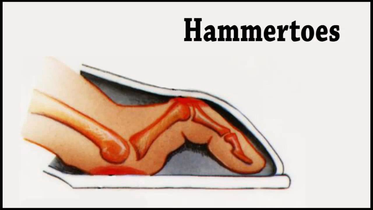
Hammer Toes Explanation
 Overview
Uneven muscle tension results in the distortion of one or several of the small toes. (Hammer toes) Pressure points develop at the raised middle joint as well as at the tip of the toe and underneath the metatarsal head. In the beginning, when the misalignment can still be corrected, it often suffices to lengthen the tendon and to cut a notch into the capsule. In a contracted misalignment, part of the middle joint is removed to form a replacement joint. Modern surgical techniques preserve the metatarsophalangeal joint (Weil or Helal osteotomies).
Causes
The cause of hammertoes varies, but causes include genetics, arthritis and injury to the toe. Treatment for hammertoes depends on the severity and can include anti-inflammatory medication, metatarsal pads, foot exercises and better-fitting shoes. If the pain caused by a hammertoe is so severe that wearing a shoe is uncomfortable, surgery may be necessary. Typically this surgery is an outpatient procedure that doesn?t require general anesthesia, though it is an option. Recovery from surgery usually takes a few weeks, and patients are given special shoes to wear.
 Symptoms
The most obvious symptom of hammertoe is the bent, hammer-like or claw-like appearance of one or more of your toes. Typically, the proximal joint of a toe will be bending upward and the distal joint will be bending downward. In some cases, both joints may bend downward, causing the toes to curl under the foot. In the variation of mallet toe, only the distal joint bends downward. Other symptoms may include Pain and stiffness during movement of the toe, Painful corns on the tops of the toe or toes from rubbing against the top of Hammer toes the shoe's toe box, Painful calluses on the bottoms of the toe or toes, Pain on the bottom of the ball of the foot, Redness and swelling at the joints. If you have any of these symptoms, especially the hammer shape, pain or stiffness in a toe or toes, you should consider consulting your physician. Even if you're not significantly bothered by some of these symptoms, the severity of a hammertoe can become worse over time and should be treated as soon as possible. Up to a point hammertoes can be treated without surgery and should be taken care of before they pass that point. After that, surgery may be the only solution.
Diagnosis
Your doctor is very likely to be able to diagnose your hammertoe simply by examining your foot. Even before that, he or she will probably ask about your family and personal medical history and evaluate your gait as you walk and the types of shoes you wear. You'll be asked about your symptoms, when they started and when they occur. You may also be asked to flex your toe so that your doctor can get an idea of your range of motion. He or she may order x-rays in order to better define your deformity.
Non Surgical Treatment
Conservative treatment is the first choice, often starting with a change of shoes to ones that have soft, larger toe spaces. Toe exercises may be prescribed to stretch and strengthen the toe muscles. Over-the-counter straps, cushions or non-medicated corn pads may be recommended to help relieve your symptoms.
Surgical Treatment
Bone-mending procedures realign the contracted toe by removing the entire deviated small joints of the toe (again, not at the ball of the foot). This allows for the buckled joint to be positioned flat and the bone ends to mend together. Often surgical hardware (fixation) is necessary to keep the bones steady during healing. Hardware options can involve a buried implant inside the toe, or a temporary wire that is removed at a later date. Medical terminology for this procedure is called a proximal interphalangeal joint arthrodesis (fusion), or a distal interphalangeal joint arthrodesis (fusion), with the former being performed in a high majority of cases.
 Prevention
There are several things you can do to help prevent hammer toes from forming or progressing. Wear supportive shoes to help prevent deformities. Hammer toes are often related to faulty foot mechanics, especially foot flattening. Wear custom orthotics prescribed by your podiatrist. Orthotics may slow the progression or prevent the development of hammer toes. Avoid shoes with narrow or pointed toe boxes that can compress the toes.
Natural Hammer Toe Treatment
 Overview
Hammer toe and mallet toe are two foot deformities that occur most often in women who wear high heels or shoes with a narrow toe box. These types of footwear may force your toes against the front of the shoe, causing an unnatural bending. A hammertoe has an abnormal bend in the middle joint of a toe. Mallet toe affects the joint nearest the toenail. Hammertoe and mallet toe are most likely to occur in the toe next to your big toe. Relieving the pain and pressure of hammertoe and mallet toe may involve changing your footwear and wearing shoe inserts. If you have a more severe case of hammertoe or mallet toe, you may need surgery to experience relief.
Causes
Medical problems, such as stroke or diabetes that affect the nerves, may also lead to hammertoe. For example, diabetes can result in poor circulation, especially in the feet. As a result, the person may not feel that their toes are bent into unnatural positions. The likelihood of developing hammertoe increases with age and may be affected by gender (more common in women) and toe length; for example, when the second toe is longer than the big toe, hammertoe is more likely to occur. Hammertoe may also be present at birth. Genetics may factor in to developing hammertoe, particularly if the foot is flat or has a high arch, resulting in instability.
 Symptoms
Signs and symptoms of hammertoe and mallet toe may include a hammer-like or claw-like appearance of a toe. In mallet toe, a deformity at the end of the toe, giving the toe a mallet-like appearance. Pain and difficulty moving the toe. Corns and calluses resulting from the toe rubbing against the inside of your footwear. Both hammertoe and mallet toe can cause pain with walking and other foot movements.
Diagnosis
Hammertoes are progressive, they don?t go away by themselves and usually they will get worse over time. However, not all cases are alike, some hammertoes progress more rapidly than others. Once your foot and ankle surgeon has evaluated your hammertoes, a treatment plan can be developed that is suited to your needs.
Non Surgical Treatment
Treating hammertoe involves straightening the toe, making tendons in the toes flexible again, and preventing the problem from returning. Some simple treatments include splinting the toe to keep it straight and to stretch the tendons of the foot. Using over-the-counter pads, cushions or straps to decrease discomfort Exercising the toes to relax the foot tendons (a session with a physical therapist may help you get started with foot exercises) Wearing shoes that hammertoes fit properly and allow toes plenty of room to stretch out.
Surgical Treatment
The deformity is corrected in a variety of ways. There are actually a large number of procedures. The simplest procedure would involve a Tenotomy, the cutting of the tendon causing the deformity or a Tendon Lengthening procedure. These procedures are infrequently done, though, as the structural deformity (the arthritis and joint adaptation) is not addressed with these surgeries. Other soft-tissue procedures involve rebalancing the tendons around the joint. There are several techniques to do this, but the most common is probably the Girdlestone-Taylor procedure, which involves rerouting the tendons on the bottom of the toe up and over the toe where it sticks up, so that the tendon helps pull the toe downwards into proper alignment.
Why Do I Have Bunions?
Overview
 A bunion is a painful deformity of the joint where the bones of the foot and the big toe meet. The enlargement of the bone and tissue around this joint is known as a bunion or hallux valgus. Symptoms of a bunion include a swollen bursal sac, a bony deformity on the side of the great toe joint, tender and swollen tissues surrounding the deformity, and displacement of the big toe, which may turn inward.
Causes
Essentially, bunions are caused by a disruption of the normal interworking of the bones, muscles, ligaments and tendons that comprise your feet, often from wearing shoes that squeeze the toes or place too much weight-bearing stress on them. However, it should be pointed out that other causes or factors in the development of bunions can include flat feet or low arches in the feet, some forms of arthritis, problems with foot mechanics, foot injuries and neuromuscular disorders such as cerebral palsy. Arthritis in the MTP joint, for example, can degrade the cartilage that protects it, and other problems may cause ligaments to become loose. Pronation, walking in a way that your foot rolls inwards, increases your risk for developing bunions.
Symptoms
Bunions may or may not cause symptoms. A frequent symptom is foot pain in the involved area when walking or wearing shoes that is relieved by resting. A bunion causes enlargement of the base of the big toe and is usually associated with positioning of the big toe toward the smaller toes. This leads to intermittent or chronic pain at the base of the big toe. Bunions that cause marked pain are often associated with swelling of the soft tissues, redness, and local tenderness. It is important to note that, in post-pubertal men and post-menopausal women, pain at the base of the big toe can be caused by gout and gouty arthritis that is similar to the pain caused by bunions.
Diagnosis
Generally, observation is adequate to diagnose a bunion, as the bump is obvious on the side of the foot or base of the big toe. However, your physician may order X-rays that will show the extent of the deformity of the foot.
Non Surgical Treatment
Technically, you can only ?fix a bunion? with surgery, but many patients don't need it to get symptom relief. In its early stages, the progression of the bunion deformity can often be dramatically slowed. Removing pressure from the bunion area and balancing the tendon and ligament alignment are the primary goals of mild bunion treatment. For example, it is important to wear shoes that have sufficient room in the toe area to accommodate the bunion - that means softer leather shoes to mold to the deformity and platform type heels for better foot and arch support. Your doctor may also advise the use of pads to protect the bunion from shoe pressure. Customized shoe inserts, called orthotics are made exclusively for your foot and are often used to correct the alignment of the arch and big toe joint. In some cases, physicians also use anti-inflammatory creams around the bunion. 
Surgical Treatment
The aim of surgery is to correct the cause of the bunion and prevent it growing back. Which type of surgery your podiatric surgeon recommends will depend on the severity of your bunion. Because there are risks and complications with any type of surgery, it?s not usually advised unless your bunions are causing pain, or if it is starting to deform your other toes.
What Actually Causes Feet To Over Pronate
Overview
When the ankles over-pronate, it throws everything out of alignment. The bones in the feet can actually shift over time, and the muscles and tendons in the leg and ankle will twist. This places repetitive stress on the ligaments, tendons, joints and muscles of the foot, which can cause injury. If your feet are over-pronating, you may notice your muscles feet unusually tired when you way or stand, or they may ache. But symptoms will vary by your weight, age and activity.
Causes
Unless there is a severe, acute injury, overpronation develops as a gradual biomechanical distortion. Several factors contribute to developing overpronation, including tibialis posterior weakness, ligament weakness, excess weight, pes planus (flat foot), genu valgum (knock knees), subtalar eversion, or other biomechanical distortions in the foot or ankle. Tibialis posterior weakness is one of the primary factors leading to overpronation. Pronation primarily is controlled by the architecture of the foot and eccentric activation of the tibialis posterior. If the tibialis posterior is weak, the muscle cannot adequately slow the natural pronation cycle.
Symptoms
With over pronation, sufferers are most likely to experience pain through the arch of the foot. A lack of stability is also a common complaint. Over pronation also causes the foot to turn outward during movement at the ankle, causing sufferers to walk along the inner portion of the foot. This not only can deliver serious pain through the heel and ankle, but it can also be the cause of pain in the knees or lower back as well. This condition also causes the arch to sink which places stress on the bones, ligaments, and tendons throughout the foot. This may yield other common conditions of foot pain such as plantar fasciitis and heel spurs.
Diagnosis
A quick way to see if you over-pronate is to look for these signs. While standing straight with bare feet on the floor, look so see if the inside of your arch or sole touches the floor. Take a look at your hiking or running shoes; look for wear on the inside of the sole. Wet your feet and walk on a surface that will show the foot mark. If you have a neutral foot you should see your heel connected to the ball of your foot by a mark roughly half of width of your sole. If you over-pronate you will see greater than half and up to the full width of your sole.
Non Surgical Treatment
Adequate footwear can often help with conditions related to flat feet and high arches. Certified Pedorthists recommend selecting shoes featuring heel counters that make the heel of the shoe stronger to help resist or reduce excessive rearfoot motions. The heel counter is the hard piece in the back of the shoe that controls the foot?s motion from side-to-side. You can quickly test the effectiveness of a shoe?s heel counter by placing the shoe in the palm of your hand and putting your thumb in the mid-portion of the heel, trying to bend the back of the shoe. A heel counter that does not bend very much will provide superior motion control. Appropriate midsole density, the firmer the density, the more it will resist motion (important for a foot that overpronates or is pes planus), and the softer the density, the more it will shock absorb (important for a cavus foot with poor shock absorption) Wide base of support through the midfoot, to provide more support under a foot that is overpronated or the middle of the foot is collapsed inward.
Prevention
Massage and stretch the calves to increase dorsiflexion at the foot/ankle. Dorsiflexion is the bending at the ankle. By improving the dorsiflexion, one will have more flexibility at the ankle, which will allow the foot to over-pronate less. Massage the IT Band with a foam roller or tennis ball to quiet down the tightness throughout this part of the leg. The IT Band attaches from the glute maximus and runs down the side of the leg into the knee area. When the IT Band is tight it will accelerate the force of the leg moving inward, which will cause the foot to move inward as well. It is often that tightness through the IT Band that promotes over-pronation. Decreasing over-pronation, which is very prominent in runners, will help add endurance, speed and efficiency to your run and ultimately place less stress on your body.
What Are The Causes Of Severs Disease?
Overview
Sever's disease occurs in children when the growth plate (which is the growing part of the heel) is injured. The foot is one of the first body parts to grow to full size. This usually occurs in early puberty. During this time, bones often grow faster than muscles and tendons. As a result, muscles and tendons become tight. The heel area is less flexible. During weight-bearing activity (activity performed while standing), the tight heel tendons may put too much pressure at the back of the heel (where the Achilles tendon attaches). This can injure the heel and cause Sever's disease.
Causes
One of the most common causes of heel pain in adolescents, Sever's disease is an overuse injury to the growth plate of the calcaneus (heel). It is commonly seen in growing, active children 8-12 years of age. Soccer players, gymnasts, and children who do any sort of running or jumping activity have an increased risk of developing Sever's disease. Occurring in one or both feet, Sever?s disease occurs at the back of the heel (also known as the apophysis or the insertion of the Achilles tendon).
Symptoms
The main symptom of sever's disease is pain and tenderness at the back of the heel which is made worse with physical activity. Tenderness will be felt especially if you press in or give the back of the heel a squeeze from the sides. There may be a lump over the painful area. Another sign is tight calf muscles resulting with reduced range of motion at the ankle. Pain may go away after a period of rest from sporting activities only to return when the young person goes back to training.
Diagnosis
Your Podiatrist or Physiotherapist will assist in diagnosing the injury and the extent of the damage. From this, they will develop a management plan which may include rest or activity modification, soft tissue treatment such as massage and stretching, correction of biomechanics through heel raises or orthoses and the progression through a series of specific strengthening exercises.
Non Surgical Treatment
Heel pain, unlike the heel spurs, that occur in adults is very uncommon in children. Of those children who do get heel pain, by far the most common cause is a disturbance to the growing area at the back of the heel bone (calcaneus) where the strong achilles tendon attaches to it. This is known as Sever's disease or calcaneal apophysitis (inflammation of the growth plate). It is most common between the ages of 10 to 14 years of age. These are one of several different 'osteochondroses' that can occur in other parts of the body, such as at the knee (Osgood-Schlatters Disease).